Sunday, April 18, 2010

Gram Stain

Gram stain of mixed Stap aureus (Gram positive cocci) and E. coli (Gram negative bacilli)


The Gram staining method, named after the Danish bacteriologist who originally devised it in 1882 (published 1884), Hans Christian Gram, is one of the most important staining techniques in microbiology. It is almost always the first test performed for the identification of bacteria. The primary stain of the Gram's method is crystal violet. Crystal violet is sometimes substituted with methylene blue, which is equally effective. The microorganisms that retain the crystal violet-iodine complex appear purple brown under microscopic examination. These microorganisms that are stained by the Gram's method are commonly classified as Gram-positive or Gram non-negative. Others that are not stained by crystal violet are referred to as Gram negative, and appear red.

Gram staining is based on the ability of bacteria cell wall to retaining the crystal violet dye during solvent treatment. The cell walls for Gram-positive microorganisms have a higher peptidoglycan and lower lipid content than gram-negative bacteria. Bacteria cell walls are stained by the crystal violet. Iodine is subsequently added as a mordant to form the crystal violet-iodine complex so that the dye cannot be removed easily. This step is commonly referred to as fixing the dye. However, subsequent treatment with a decolorizer, which is a mixed solvent of ethanol and acetone, dissolves the lipid layer from the gram-negative cells. The removal of the lipid layer enhances the leaching of the primary stain from the cells into the surrounding solvent. In contrast, the solvent dehydrates the thicker Gram-positive cell walls, closing the pores as the cell wall shrinks during dehydration. As a result, the diffusion of the violet-iodine complex is blocked, and the bacteria remain stained. The length of the decolorization is critical in differentiating the gram-positive bacteria from the gram-negative bacteria. A prolonged exposure to the decolorizing agent will remove all the stain from both types of bacteria. Some Gram-positive bacteria may lose the stain easily and therefore appear as a mixture of Gram-positive and Gram-negative bacteria (Gram-variable).

Finally, a counterstain of basic fuchsin is applied to the smear to give decolorized gram-negative bacteria a pink color. Some laboratories use safranin as a counterstain instead. Basic fuchsin stains many Gram-negative bacteria more intensely than does safranin, making them easier to see. Some bacteria which are poorly stained by safranin, such as Haemophilus spp., Legionella spp., and some anaerobic bacteria, are readily stained by basic fuchsin, but not safranin. The polychromatic nature of the gram stain enables determination of the size and shape of both Gram-negative and Gram-positive bacteria. If desired, the slides can be permanently mounted and preserved for record keeping.

Besides Gram's stain, there are a wide range of other staining methods available. By using appropriate dyes, different parts of the bacteria structures such as capsules, flagella, granules, and spores can be stained. Staining techniques are widely used to visualize those components that are otherwise too difficult to see under a light microscope. In addition, special stains can be used to visualize other microorganisms not readily visualized by the Gram stain, such as mycobacteria, rickettsia, spirochetes, and others. In addition, there are modifications of the Gram stain that allow morphologic analysis of eukaryotic cells in clinical specimens.


Monday, April 12, 2010

James Lind and Scurvy

Lind was a Scottish doctor, a pioneer of naval hygiene and expert on the treatment of scurvy. . By conducting the first ever clinical trial,  he developed the theory that citrus fruits cured scurvy

James Lind was born in Edinburgh in 1716. In 1731, he registered as an apprentice at the College of Surgeons in Edinburgh and in 1739 became a surgeon's mate, seeing service in the Mediterranean, Guinea and the West Indies, as well as the English Channel. In 1747, while serving as surgeon on HMS Salisbury, he carried out experiments to discover the cause of scurvy, the symptoms of which included loose teeth, bleeding gums and haemorrhages.

Lind selected 12 men from the ship, all suffering from scurvy, and divided them into six pairs, giving each group different additions to their basic diet. Some were given cider, others seawater, others a mixture of garlic, mustard and horseradish. Another group of two were given spoonfuls of vinegar, and the last two oranges and lemons. Those fed citrus fruits experienced a remarkable recovery. While there was nothing new about his discovery - the benefits of lime juice had been known for centuries - Lind had definitively established the superiority of citrus fruits above all other 'remedies'.

In 1748, Lind retired from the navy and went to Edinburgh University to take professional qualifications. In 1753, he published 'A Treatise of the Scurvy' and in 1757 'An Essay on the Most Effectual Means of Preserving the Health of Seamen in the Royal Navy', which threw much light on the appalling living conditions and diet of seamen. In 1758, he was appointed physician to the Naval Hospital at Haslar in Gosport where he investigated the distillation of fresh water from salt water for supply to ships.

In 1763, Lind published work on typhus fever in ships and in the 1768 publication 'An Essay on Diseases Incidental to Europeans in Hot Climates' he summarised the prevalent diseases in each colony and gave advice on avoiding tropical infections. Lind died in 1794 in Gosport.

Although the importance of Lind's findings on scurvy were recognised at the time, it was not until more than 40 years later that an official Admiralty order was issued on the supply of lemon juice to ships. With this, scurvy disappeared almost completely from the Royal Navy.

Blood groups

Marcello Malpighi

Malpighi was born in Crevalcore near Bologna in Italy, raised on the farm his parents owned and entered the University of Bologna at the age of 17. Malpighi began to study. When his father, mother and paternal grandmother died, he had to abandon his studies for more than two years to settle family affairs. He returned to university after two years, and became a doctor of medicine in 1653. The next year he married Francesca Massari, younger sister of his anatomy professor. She died a year later.

In 1656 Malpighi received a chair of medical practice in the university, three years after he had applied for it, and later the same year University of Pisa created a chair of theoretical medicine for him. He stayed in Pisa for three years and then returned to Bologna. In 1661 he was called to University of Messina where he stayed for four years.


Most of Malpighi's research results were published as articles in the journal of the Royal Society of England. His first article appeared there in 1661 and was about anatomy of a lung of a frog during which he had discovered capillaries. In 1667 Henry Oldenburg invited Malpighi to correspond with the Royal Society regularly and he became a fellow the next year, the first such recognition given to an Italian.

Malpighi used the microscope for studies on skin, kidney, and for the first interspecies comparison of the liver. He greatly extended the science of embryology. The use of microscopes enabled him to describe the development of the chick in its egg, and discovered that insects (particularly, the silk worm) do not use lungs to breathe, but small holes in their skin called tracheae. Later he falsely concluded that plants had similar tubules. However, he observed that when a ringlike portion of bark was removed on a trunk a swelling of the tissues would occur above the ring. He correctly interpreted this as growth stimulated by food coming down from the leaves, and being blocked above the ring. He was the first to see capillaries and discovered the link between arteries and veins that had eluded William Harvey.

Malpighi is regarded as the founder of microscopic anatomy and the first histologist. Many microscopic anatomical structures are named after him, including a skin layer (Malpighi layer) and two different Malpighian corpuscles in the kidneys and the spleen, as well as the Malpighian tubules in the excretory system of insects.

He also studied chick embryo development with detailed drawings and discovered taste buds of human tongue. Some of his studies he made by vivisection. He also studied the anatomy of a brain and concluded that this organ is a gland. In terms of modern endocrinology this deduction is correct because neurotransmitter substances represent paracrine hormones, and the hypothalamus of the brain has long been recognized for its hormone-secreting capacity. He was also among the first to study human fingerprints.

In 1691 Pope Innocent XII invited him to Rome as Papal physician. He taught medicine in the Papal Medical School and wrote a long treatise about his studies which he donated to the Royal Society of London.

Marcello Malpighi died of apoplexy in Rome on September 30, 1694

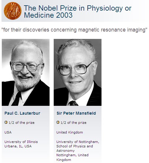
MRI - Nobel Prize

Prions and Prusiner

                                  
The word prion, coined in 1982 by Dr. Stanley B. Prusiner, is a portmanteau derived from the words protein and infectious.

Stanley Ben Prusiner (born May 28, 1942[1]) is an American neurologist and biochemist. Currently the director of the Institute for Neurodegenerative Diseases at University of California, San Francisco (UCSF). Prusiner discovered prions, a class of infectious self-reproducing pathogens primarily or solely composed of protein. He received the Albert Lasker Award for Basic Medical Research in 1994 and the Nobel Prize in Physiology or Medicine in 1997 for his prion research.

Warren and Marshall

Two Australian scientists who upset medical dogma by discovering a bacterium that causes stomach inflammation, ulcers and cancer won the 2005 Nobel Prize for Physiology or Medicine. The findings by the Australians in the early 1980's went so against medical thinking, which held that psychological stress caused stomach and duodenal ulcers, that it took many more years for an entrenched medical profession to accept it.

In the early 1980's, Dr. Warren noted the bacterium in the lower part of the stomach in about half of the patients who had biopsies. He made a crucial observation that signs of inflammation were always present in the surface lining of the stomach near where he observed the bacterium.


Dr. Marshall joined Dr. Warren in studying biopsies from a series of patients. After several attempts, Dr. Marshall succeeded in growing a bacterium that was unknown then; he named it Campylobacter pyloridis, believing that it was a member of the Campylobacter family. (It was later found to be a member of the Helicobacter family and renamed H. pylori.)

Still, many doctors were unconvinced by the findings, a point recognized by the Nobel committee, which said the award went to Dr. Marshall and Dr. Warren "who with tenacity and a prepared mind challenged prevailing dogmas."

Dr. Marshall carried on a medical tradition in experimenting on himself to test his and Dr. Warren's theory and to show that Helicobacter was the primary cause of gastritis, not a secondary invader.

In earlier interviews, Dr. Marshall described how at age 32, he swallowed a gastroscope tube to allow another doctor to look at his stomach and take several biopsies. These procedures and examinations were needed to document that Dr. Marshall had no H. pylori in his stomach and did not suffer from gastritis or another abnormality.

Dr. Marshall waited 10 days for the areas that had been biopsied to heal and then swallowed a pure culture of H. pylori. A week later, he had an unusual sensation of fullness after eating supper and felt ill. Friends told him that his breath was "putrid."

Ten days after the onset of symptoms, Dr. Marshall underwent the first of an additional three gastroscopies. Biopsies obtained through them showed that he had developed gastritis or inflammation of the stomach, but he did not continue the experiment long enough to develop an ulcer. His symptoms quickly disappeared after treatment.
Source:http://www.nytimes.com/2005/10/04/science/04nobe.html?pagewanted=2